New Q fever flyers in Portuguese/ Novos folhetos sobre a febre Q em português
Three new flyers providing information about Q fever and highlighting important precautionary measures are now available in Portuguese. The materials are aimed at different target groups:
Estão agora disponíveis três novos folhetos em português que fornecem informações sobre a febre Q e salientam medidas de precaução importantes. Os materiais destinam-se a diferentes grupos-alvo:
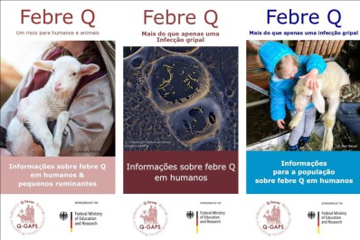
Flyer for Animal Owners & Veterinarians/Folheto para veterinários e donos de animais:
Q Fever – A Risk for Humans and Animals/Febre Q – Um Risco para Humanos e Animais
Flyer for medical doctors/Folheto para Médicos:
Q fever - More than a flu /Febre Q – Mais do que apenas uma Infecção gripal
Flyer for the general public/Folheto para o público:
Q fever - More than a flu/Febre Q – Mais do que apenas uma Infecção gripal
Flyers have already been produced in English and French as part of the Q-GAPS project. With these new Portuguese versions, we aim to make important health information accessible to an even wider international audience.
No âmbito do projeto Q-GAPS, já foram produzidos folhetos em inglês e francês. Com as novas versões em português, pretendemos contribuir para tornar informações importantes sobre saúde mais acessíveis a um público internacional mais vasto.
Download Flyer for Animal Owners & Veterinarians/Folheto para veterinários e donos de animais
Download Flyer for medical doctors/Folheto para Médicos
Download Flyer for the general public/Folheto para o público
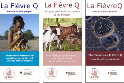
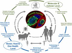 Fig. 1. Schematic representation of tasks, goals, and networking of the interdisciplinary Q-GAPS (Q fever GermAn interdisciplinary Program for reSearch) consortium. Q-GAPS is a multidisciplinary network of scientists dedicated to implementing the One Health approach. Q-GAPS aims to support the public health and veterinary communities in preventing and controlling Q fever.